
My pet is coming in for a COHAT. What does that mean?
- Audubon Family Vets

- Oct 31, 2023
- 5 min read

C.O.H.A.T. stands for Comprehensive Oral Health Assessment and Treatment.
This procedure, which is more commonly known as a dental cleaning, has many benefits for your pet. Periodontal disease is one of the most common diseases that affects dogs and cats.
Most dental issues cannot be assessed without an in-depth examination under anesthesia by your veterinarian. Pets typically don’t stop eating or show obvious signs of discomfort due to dental disease. In fact, the most common complaint from owners is that they have bad breath! That bad breath is a BIG sign that there is dental disease. Halitosis (bad breath) most commonly arises from a combination of bacteria and tartar build-up in the mouth (see below).
What is tartar?
After a person or pet eats, plaque begins to build up on their teeth. Plaque is a combination of saliva, bacteria, and food. Within 24 hours this substance begins to harden, which is why brushing your teeth (and your pet’s!) is so important. If the teeth are not brushed, plaque begins to harden, and it turns into tartar. Tartar (usually brown and yellow as seen below) can get stuck below the gumline in dogs and cats and create an ideal environment for bacteria to thrive. Subgingival tartar, which can lead to gingivitis (gum disease), is not able to be seen or accessed in an awake patient.

Once this occurs, tartar can only be removed by ultrasonic or hand-scaling during a professional dental cleaning. If this disease process goes on long enough, the inflammation from the tartar and bacteria can start to erode the surrounding gum tissue and bone, leading to advanced periodontal disease. The symptoms of this are mobile teeth, severe halitosis, gum recession, missing teeth, bleeding gums, etc. As if this process wasn’t painful enough (think back to any time you’ve had tooth pain), this bacteria load in the mouth can lead to consequences throughout the body and has been implicated in diseases in other organs such as the heart and kidneys.
My vet has recommended a dental cleaning. What’s next?
Prior to scheduling this procedure, your pet should have:
1. A pre-anesthetic exam to evaluate your pet’s overall health with the vet who will be performing the procedure. Your vet will palpate all the structures surrounding the mouth including the lips, the chewing muscles, the salivary glands, the lymph nodes, and the bones and tissues surrounding the eyes. All of these can be involved when there is dental disease. Your vet will also examine the teeth, tongue, gingiva, and intra-oral structures as much as possible. This can give your vet an idea about the degree of dental disease, but an anesthetic exam is still required.
2. Pre-anesthetic blood work is strongly encouraged to obtain baseline values as well as to screen for underlying health issues.
3. Sometimes, additional testing such as x-rays or an echocardiogram will be recommended if your pet is older or has a history of heart or lung disease.
4. Pre-anesthetic medications that help with sedation, anxiety, and nausea will be dispensed to help your pet as they enter the hospital on the morning of the procedure.
The Procedure
Your pet will receive a thorough pre-anesthetic exam by your veterinarian prior to anesthesia the morning of the procedure. Any preoperative testing will be completed, and any abnormalities will be discussed with you over the phone.
Your pet will then have an intravenous catheter placed to facilitate anesthetic medications and fluid therapy. Your pet will have an individually tailored anesthetic plan. They will have an endotracheal tube placed in their airway to deliver oxygen and anesthetic gasses. Not only is this vital for anesthesia, but this tube also protects the pet’s airway from accidental aspiration (when water, food, and other material goes into the trachea and airways). Your pet will have their vital signs and anesthesia monitored by a dedicated technician throughout the entire procedure.
A COHAT begins with a thorough examination of all the teeth and mouth structures under anesthesia. The teeth are probed, examined, and any abnormalities are noted in the patient’s chart. Next, all teeth are scaled, cleaned, and polished.

X-rays of each tooth and its root(s) are taken to evaluate for underlying disease. Lastly, your veterinarian determines a treatment plan for any diseased teeth or abnormalities in the mouth. This can involve extractions of diseased teeth, removal of masses or gingival overgrowth, or consult with a board-certified veterinary dentist. Pets that require extractions receive local anesthetic (similar to Novocain in people) as well as pain medications during and after surgery.
Teeth are like icebergs. What we can see on the surface is sometimes vastly different from disease that lives beneath the gum line.

A Happy Ending
Molly, a sweet Dachshund, was brought to us because her owners suspected that she had periodontal disease. They noted bad breath, discolored teeth, and she had a tooth root abscess. Her owners were very diligent and wanted to take care of her tooth pain as soon as possible. We saw gingivitis, tartar, and a couple of mobile teeth on her awake exam, and so we scheduled her for a COHAT.

Under anesthesia, we began cleaning her teeth and took x-rays of each tooth. We discovered that she required several tooth extractions as well as a general cleaning on the rest of her teeth.

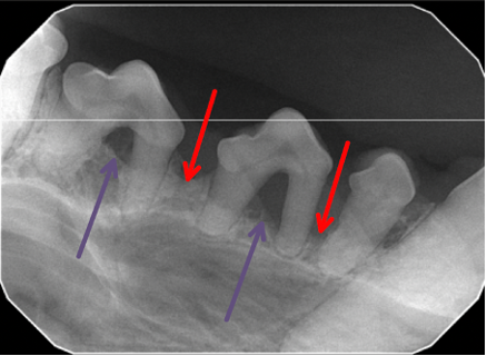
Below are Molly’s x-rays. Several of her premolars (the small teeth on the sides of the mouth behind the canine teeth) appeared normal on the surface. However, when a dental probe was gently applied, we discovered that she had periodontal pockets around these teeth. A periodontal pocket means that the gum tissue and often bone surrounding the tooth have detached from the tooth deeper than they should, indicating advanced periodontal disease. She also had furcation exposure (visible gap between the roots). These teeth were loose and painful without the proper support of the bone and gingiva.
Compare the x-ray on the left (normal) with the one on the right (abnormal). The bone surrounding the teeth (red arrows) should be level with the crown (the visible portion of the tooth on the surface). Note that the bone on the right has decreased almost to the tip/point of the tooth roots in the diseased teeth. The area between the roots of a tooth is called a furcation. Normally, this should be supported by bone. Note that the purple arrows on the left demonstrate adequate bone coverage of the furcation, while the purple arrows on the right demonstrate exposed, painful root furcations. This would not have been visible without x-rays.


Molly’s teeth were thoroughly cleaned, and the diseased teeth were removed. Her gingiva was stitched up and she was put on a wet food diet for the next couple weeks while her mouth healed. Her mouth was now sparkly clean and pain-free!
Pets do incredibly well even with few teeth due to the way they chew their food. Most pets can still chew on bones, eat hard food, and realistically go about their normal life after tooth removal. We would rather a pet have a pain-free mouth with less teeth than a painful mouth full of teeth. Owners often report that their dogs eat better the night after surgery even with stitches (because the painful teeth are removed) than they ate prior! Better yet, regular cleanings can PREVENT periodontal disease and lessen the chance that any teeth will need to be removed. In summary, COHATs provide an incredible benefit to your pet.
For further reading:
- Pet periodontal disease: https://afd.avdc.org/pet-periodontal-disease/
- Stages of pet periodontal disease: https://afd.avdc.org/five-stages-of-pet-periodontal-disease/
- Questions to ask your vet about pet dental cleanings: https://afd.avdc.org/questions-to-ask-your-veterinarian-about-pet-dental-cleanings/
- AVDC COHAT explanation: https://afd.avdc.org/what-is-a-professional-veterinary-dental-cleaning/
- What is an anesthesia-free dental cleaning?https://afd.avdc.org/what-is-an-anesthesia-free-dental-cleaning/


Comments